38 the human heart and its labels
Label the heart - Science Learning Hub Label the heart Add to collection In this interactive, you can label parts of the human heart. Drag and drop the text labels onto the boxes next to the diagram. Selecting or hovering over a box will highlight each area in the diagram. Pulmonary vein Right atrium Semilunar valve Left ventricle Vena cava Right ventricle Pulmonary artery Aorta Given alongside is a diagram of the human heart showing ... Given alongside is a diagram of the human heart showing its internal structure. Label the parts marked 1 to 6, and answer the following questions.Which chamb...
Heart Anatomy: Labeled Diagram, Structures, Blood Flow ... Image: Use the 2x2 table to label the 4 chambers of the heart, including the right atrium, right ventricle, left atrium, and left ventricle. Tricuspid Valve and Mitral Valve Now that we have a good understanding of the 4 chambers of the heart, let's move on to the 4 main valves.

The human heart and its labels
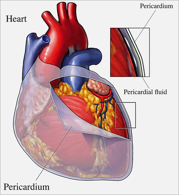
Human Genome Project Results Nov 12, 2018 · By using mRNA as a template, scientists use enzymatic reactions to convert its information back into cDNA and then clone it, creating a collection of cDNAs, or a cDNA library. These libraries are important to scientists because they consist of clones of all protein-encoding DNA, or all of the genes, in the human genome. The Anatomy of the Heart, Its Structures, and Functions The heart is the organ that helps supply blood and oxygen to all parts of the body. It is divided by a partition (or septum) into two halves. The halves are, in turn, divided into four chambers. The heart is situated within the chest cavity and surrounded by a fluid-filled sac called the pericardium. This amazing muscle produces electrical ... Heart Diagram – 15+ Free Printable Word, Excel, EPS, PSD ... Teachers and students use the heart diagram, in biological science, to study the structure and functions of a human being’s heart. Friends and colleagues on the other hand may find this diagram template useful when it comes to sending special, personalized gifts to their family members and significant others. Download the template today, and ...
The human heart and its labels. A Labeled Diagram of the Human Heart You Really Need to ... The human heart, comprises four chambers: right atrium, left atrium, right ventricle and left ventricle. The two upper chambers are called the left and the right atria, and the two lower chambers are known as the left and the right ventricles. The two atria and ventricles are separated from each other by a muscle wall called 'septum'. Heart Diagram with Labels and Detailed Explanation - BYJUS The human heart is the most crucial organ of the human body. It pumps blood from the heart to different parts of the body and back to the heart. The most common heart attack symptoms or warning signs are chest pain, breathlessness, nausea, sweating etc. The diagram of heart is beneficial for Class 10 and 12 and is frequently asked in the ... A Diagram of the Heart and Its Functioning Explained in ... The heart blood flow diagram (flowchart) given below will help you to understand the pathway of blood through the heart.Initial five points denotes impure or deoxygenated blood and the last five points denotes pure or oxygenated blood. 1.Different Parts of the Body ↓ 2.Major Veins ↓ 3.Right Atrium ↓ 4.Right Ventricle ↓ 5.Pulmonary Artery ↓ 6.Lungs Parts Of The Human Heart - Science Trends The Anatomy of the Human Heart. Here are some important facts about the anatomy of the human heart: The human heart can weight between 200 to 425 grams (or 7 and 15 ounces). On average it beats about 100,000 times a day and pumps 7,571 liters (2,000 gallons) of blood.
File:Diagram of the human heart (cropped).svg - Wikimedia Apr 05, 2022 · Add Inferior vena cava and pericardium labels: 18:08, 14 August 2018: 656 × 631 (209 KB) ... Diagram of the human heart, created by Wapcaplet in Sodipodi. Cropped by ... How to Draw the Internal Structure of the Heart (with ... Make sure to label the following: Superior Vena Cava Inferior Vena Cava Pulmonary Artery Pulmonary Veins Left Ventricle Right Ventricle Left Atrium Right Atrium Mitral Valves Aortic Valves Aorta Pulmonic Valve (Optional) Tricuspid Valve (Optional) 6 To finish, label "The Human Heart" above the sketch. Community Q&A Search Add New Question Question Normal chest MDCT with anatomic labels | e-Anatomy - IMAIOS Mar 10, 2022 · Pocket Atlas of Human Anatomy: 5th edition - W. Dauber, Founded by Heinz Fene Anatomical variants and notes from the author about the anatomical labeling of the thorax CT: In the lower lobe of the left lung, there is an inconstant subsuperior pulmonary segment that is seen in approximately 30% of individuals, located between the superior and ... Heart: illustrated anatomy - e-Anatomy This interactive atlas of human heart anatomy is based on medical illustrations and cadaver photography. The user can show or hide the anatomical labels which provide a useful tool to create illustrations perfectly adapted for teaching. Anatomy of the heart: anatomical illustrations and structures, 3D model and photographs of dissection.
Heart: Anatomy and Function Your heart is the main organ of your cardiovascular system, a network of blood vessels that pumps blood throughout your body. It also works with other body systems to control your heart rate and blood pressure. Your family history, personal health history and lifestyle all affect how well your heart works. Appointments 800.659.7822 Human Heart (Anatomy): Diagram, Function, Chambers ... The heart is a muscular organ about the size of a fist, located just behind and slightly left of the breastbone. The heart pumps blood through the network of arteries and veins called the... Human Heart Diagram Labeled - Science Trends Human Heart Diagram Labeled Daniel Nelson 1, January 2019 | Last Updated: 3, March 2020 The human heart is an organ responsible for pumping blood through the body, moving the blood (which carries valuable oxygen) to all the tissues in the body. Without the heart, the tissues couldn't get the oxygen they need and would die. File:Diagram of the human heart (no labels).svg ... File:Diagram of the human heart (no labels).svg. Size of this PNG preview of this SVG file: 498 × 599 pixels. Other resolutions: 199 × 240 pixels | 399 × 480 pixels | 499 × 600 pixels | 639 × 768 pixels | 851 × 1,024 pixels | 1,703 × 2,048 pixels | 533 × 641 pixels.
Heart - Wikipedia The human heart is situated in the mediastinum, at the level of thoracic vertebrae T5-T8.A double-membraned sac called the pericardium surrounds the heart and attaches to the mediastinum. The back surface of the heart lies near the vertebral column, and the front surface sits behind the sternum and rib cartilages. The upper part of the heart is the attachment point for several large blood ...
Human Heart - Anatomy, Functions and Facts about Heart The human heart is about the size of a human fist and is divided into four chambers, namely two ventricles and two atria. The ventricles are the chambers that pump blood and atrium are the chambers that receive blood. Among which both right atrium and ventricle make up the "right heart," and the left atrium and ventricle make up the "left heart."
Citizen Sleeper review: a stylish machine with a gooey human ... May 03, 2022 · Throwing off the shackles of a faceless process governing your life is a recurring theme in this blend of sci-fi RPG and interactive fiction, and it makes for a strong rags-to-ramen story of one robot on the run. Even when the game's own systems of dice and clocks clash with its stories of human interest, it is the people who come out on top.
Human Heart - Diagram and Anatomy of the Heart The heart is a muscular organ about the size of a closed fist that functions as the body's circulatory pump. It takes in deoxygenated blood through the veins and delivers it to the lungs for oxygenation before pumping it into the various arteries (which provide oxygen and nutrients to body tissues by transporting the blood throughout the body).
Heart Anatomy With Labels Photos and Premium High Res ... Browse 147 heart anatomy with labels stock photos and images available, or start a new search to explore more stock photos and images. heart blood flow - heart anatomy with labels stock illustrations. human heart anatomy. blood flow - heart anatomy with labels stock illustrations. circulatory system, diagram - heart anatomy with labels stock ...
Diagram of Human Heart and Blood Circulation in It | New ... Exterior of the Human Heart A heart diagram labeled will provide plenty of information about the structure of your heart, including the wall of your heart. The wall of the heart has three different layers, such as the Myocardium, the Epicardium, and the Endocardium. Here's more about these three layers. Epicardium
The Heart - Science Quiz - Seterra The Heart - Science Quiz: Day after day, your heart beats about 100,000 times, pumping 2,000 gallons of blood through 60,000 miles of blood vessels. If one of your organs is working that hard, it makes sense to learn about how it functions! This science quiz game will help you identify the parts of the human heart with ease. Blood comes in through veins and exists via arteries—to control the ...
13 parts of the human heart (and its functions ... Although the heart is connected and is influenced by the nervous system, it actually acts largely autonomously. Parts of the heart and its functions. The human heart is shaped by different parts whose coordinated action allows the pumping of blood. It is widely known that we can find four chambers inside the heart: two atria and two ventricles.


Post a Comment for "38 the human heart and its labels"